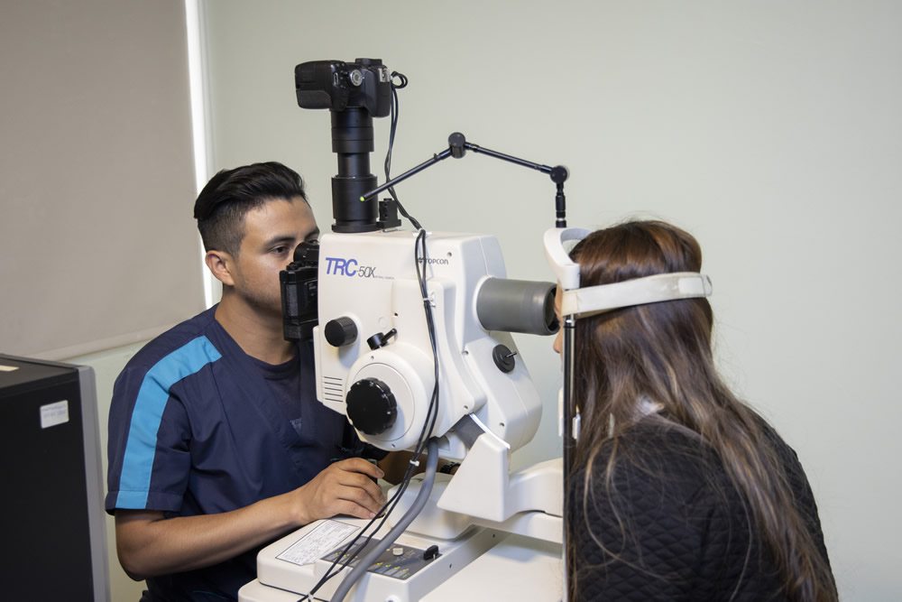
The retinal camera or study of the eye fundus is one of the diagnosis techniques focus on visualizing the posterior pole of the eye globe, this includes the retina, optic disc, choroids, and blood vessels.
There are three basic kinds of ophthalmoscopy:
- Direct ophthalmoscopy: Simple technique in which the ocular exploration is done using a monocular ophthalmoscopy.
- Indirect ophthalmoscopy: Technique in which the ocular exploration is done using a binocular ophthalmoscopy and an external light source.
- Indirect ophthalmoscopy with slit lamp: Complex technique in which the ocular exploration is done using the slit lamp for a more accurate diagnosis.
CASES IN WHICH THIS RETINAL IMAGING IS NEEDED:
Through the retinal camera and imaging, ophthalmologist can see different diseases and pathologies in the eye most important area. Here is possible to see blood vessels, the retina, and the optic nerve, three essential elements not only for the vision but as structures which alterations can be fundamental to diagnose different eye diseases.

