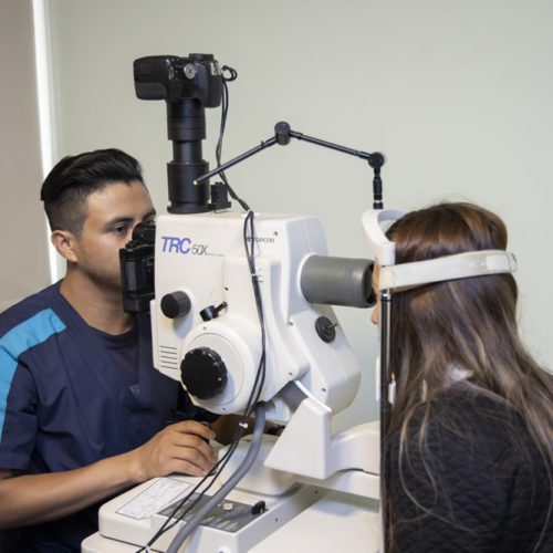
The ophthalmoscopy or study of the eye fundus is one of the diagnosis techniques focus on visualizing the posterior pole of the eye globe, this includes the retina, optic disc, choroids, and blood vessels.
Project Category: Exams
Interferometry
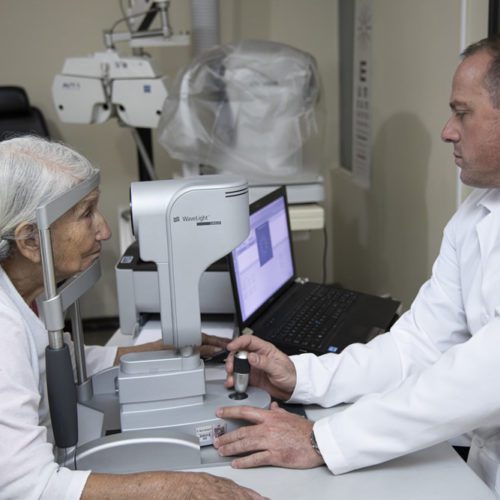
Modern equipment (lenstar) specialized to perform Intra Ocular Lenses calculation which is used for the cataract surgery.
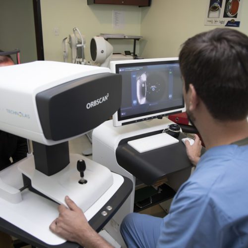
OrbScan
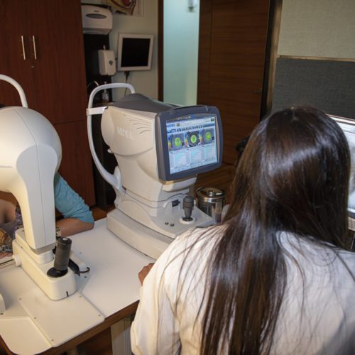
This topography provides complete information about the corneal structure and anterior chamber. The OrbScan topography measures the shape of the corneal frontal and anterior surface, this gives a complete image of the cornea thickness.
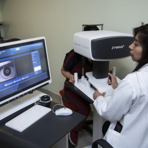
ZWave
This Aberrometer measures and analyses the presence of aberrations of high and low order. It has iris recognition for a safe and efficient identification of the patient, it also compensates the change of the pupillary axis and cyclotorsion.
OPD-Scan III
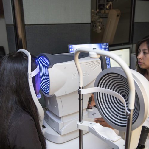
Third generation of aberrometer / corneal topographer, it allows to have wide and accurate information about the eye refractive status, providing an integral eye evaluation.
Keratograph 5M
This keratograph is an advanced corneal topographer with a real integrated keratometry and an optimized color camera for external images.
Multi-Diagnostic Platform
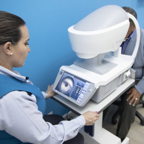
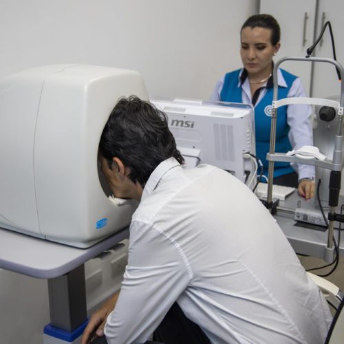
This equipment combines functions from autorefractometer, keratometry, corneal topographer, aberrometer, pachymeter and tonometer combining several features for anterior chamber analysis making it one of the most advance platforms.
Electrophysiology
Electrooculogram (EOG), Electroretinogram (ERG), Cobra Retinal Camera, Visual Evoked Potentials, Visual Training
- 1
- 2

